- e-Stroke has enabled the stroke team to reduce their door-to-groin-puncture time by 8 minutes (a 10% improvement) - an important step towards improved patient outcomes
- "The e-Stroke is absolutely advanced and helps us to provide significantly improved patient care…it stands out from the crowd because it delivers very reliable results."
The following is an English translation of an article appearing in Radiologie Magazin in March 2021 (click here to view the original article).
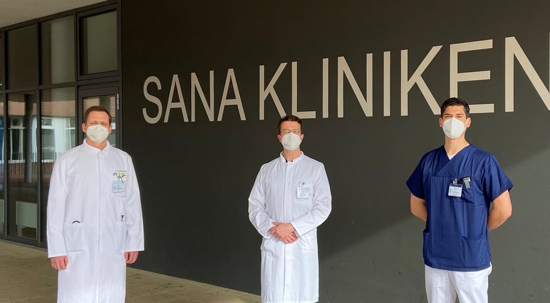
As a member of the Schleswig-Holstein South stroke network, Sana Kliniken Lübeck is known for its speedy diagnostics and experienced therapy for acute stroke. Whether it is thrombolysis or minimally invasive intracranial thrombectomy, Prof. Jan Peter Goltz (Chief Physician of the Institute for Diagnostics and Interventional Radiology and Deputy Director of the Heart & Vascular Center), Dr. Jens Schaumberg (Head of the Department of Neurology), and Dr. Christian Dyzmann (Section Head Neuroradiologist) work closely together to provide patients with the fastest possible and most therapeutically effective care.
Outside of regular working hours (i.e., nights, weekends, holidays), there is a neuroradiological on-call service for intracranial thrombectomy, as in most hospitals in Germany. Since every minute is critical in stroke, the hospital management decided to implement the AI software to improve the diagnostic workflow during and outside of regular working hours. Since February 2020 the team around Prof. Goltz, Dr. Schaumberg and Dr. Dyzmann have been using e-Stroke by Brainomix. Right from the start, the aim was to further improve stroke care, which, due to previous optimizations, was already at an above-average level.
Optimize Workflow and Shorten Treatment Times
"As there is no interventional neuroradiologist on site at the hospital at night, on weekends and during public holidays, we were looking for a solution that supports our workflow in examining patients with suspected stroke during this period", describes the neuroradiologist, Dr. Christian Dyzmann.
Before the introduction of the new software, the neuroradiologist on duty would need to dial into the hospital system via external access to view the images of the CT examination, while the treating neurologist was in contact with him on site in the hospital by phone. But before this call was made, much time may have already passed – hence the need for the new software.
Today the scans from Sana Kliniken are automatically analyzed with an AI algorithm, and those images from patients with a suspected stroke are sent to the stroke specialists immediately. This step is done automatically, with almost no time delay. The evaluation delivers reformatted brain CT images, as well as an assessment of the severity of the stroke and the level of infarction (ASPECT Score), locates the clot in the cerebral vessels, and generates a CT angiography and CT perfusion evaluation marking the occlusion within the middle cerebral artery. The hospital management agreed to invest in the AI-based decision support software, e-Stroke, from Brainomix.
The neurologist Dr. Jens Schaumberg is enthusiastic: “The advantage of the software solution from Brainomix is that both the neuroradiologists and us neurologists receive the scans simultaneously on our smartphones, which enables us to determine the most appropriate therapy much faster.” With this transmission, the specialists see the images very quickly, and can determine whether the patient is eligible for thrombolysis and/or mechanical thrombectomy. Figures shows that 10-15 percent of stroke patients may be eligible for thrombectomy. At Sana Kliniken, 23 percent of all stroke patients are treated with thrombolysis.
Prof. Jan Peter Goltz: “Compared to supportive software packages for radiology, which I got to know during my time at university, I can confirm that the e-Stroke software from Brainomix is absolutely advanced and helps us to provide significantly improved patient care. The Brainomix software solution stands out from the crowd because it delivers very reliable results."
The Evaluation is Automated
"Time is brain,” explains Dr. Dyzmann. “For stroke patients, every single minute really counts. Any support that helps to shorten the time to treatment makes perfect sense. This is why the neuroradiologists and neurologists at Sana Kliniken Lübeck evaluated the market to see which software packages have already been validated to evaluate strokes.”
Dr. Jens Schaumberg: “It used to be the case that the senior physician or neuroradiologist always had to come to the clinic for perfusion imaging in order to evaluate the data locally. That is why the e-Stroke software represents such a quantum leap for us. The earlier thromboslysis or intracranial thrombectomy can be performed, the more successful the treatment will be. The Brainomix app and e-Stroke software have significantly improved the way the Lübeck stroke team works.”
During the day, the stroke specialists are alerted by a call from the ambulance control center as soon as an acute stroke is brought to the hospital. They immediately see to it that the scanner is kept free. The neuroradiologists and neurologists wait for the patient at the CT.
“Most stroke patients are announced so that we are informed in advance of their age or the stroke symptoms. We can then still see during the examination whether the native CT scan is sufficient or whether we have to do a CT perfusion or CTA in order to detect a possible cerebral artery occlusion,” notes Dr. Jens Schaumberg.
All doctors involved have the Brainomix app on their smartphones and get the fully automated analysis of findings sent to their device via push function immediately after the examination. “During the on-call service, we no longer necessarily need to start up a computer. The new workflow greatly simplifies the service system. We are also much more flexible and even faster during our on-call service at night, on weekends and on public holidays,” reports Dr. Christian Dyzmann.
Immediately after receipt of the findings on the smartphone, he and the neurological colleague can establish the indication for an intracranial thrombectomy, call the anaesthetist and inform the radiologist. And then it basically starts. In the clinic, a coronary MIP reconstruction of the CTA is only checked for the access route so that he knows which catheters are required for intervention, or whether there are difficulties in the access route. For Dyzmann, the analysis with e-Stroke from Brainomix has been the "ground truth" for almost a year. He is enthusiastic about the evaluation and presentation of the results in the app.
More Flexibility in Service
After a brief evaluation period, the neuroradiologists came to appreciate the Brainomix app as a very helpful tool for establishing the indication for thrombectomy. They can assess the severity of the stroke and recognize the vascular occlusion in the processed MIP. Using the more sensitive and specific CT brain perfusion, Dr Dyzmann even recognizes where the blockage must be. The app provides him with exactly the information he needs to alert the interventional team.
“Compared to the time before we implemented e-Stroke, we have saved several minutes in on-call service – that is, from the arrival of the patient at the hospital to the time of groin puncture (i.e., door-to-groin), we are at eight minutes faster,” says the neuroradiologist after examining the first evaluations. “A doctoral student in our department is already analyzing the exact effects on the clinic's internal workflow,” adds Prof. Goltz.
The literature on the benefits of intracranial thrombectomies shows that ten minutes of treatment time saved means that the patient is one percent more likely to survive the stroke without being disabled. “Our expectations for the software were not only met, but exceeded,” confirm Prof. Goltz, Dr. Schaumberg and Dr. Dyzmann in unison. The system was ready for use after less than five hours of installation, and shortly afterwards the e-Stroke transmitted the first evaluation of a stroke. And the good cooperation continues to this day. All software updates are seamlessly implemented, and any changes requested by the stroke specialists are made to the software within a short period of time. Dyzmann and his colleagues rate the support from Brainomix as unparalleled.
From a technical point of view, the CT examinations are routed directly to the PACS and in parallel to the locally installed e-Stroke software. The processing time of a native skull CT takes about one minute, that of the CTA one minute and 20 seconds and that of the CT perfusion no longer than one minute and 30 seconds. The analysis software runs on virtual servers in the PACS - all patient data remains in the hospital. The images are only transferred to the app in pseudonymised form via a cloud service. The entire process is completely automated in the background. It does not require a single click, neither from the radiographer nor from a doctor.
The only wish that Prof. Goltz, Dr. Schaumberg and Dr. Dyzmann have for Brainomix: “At the moment only occlusions in the middle cerebral artery and intracranial main artery are recognized. It would be nice if the occlusions of the anterior and posterior cerebral arteries were recognized, and a reconstruction of the extracranial course of the arteries supplying the brain were made available.” However, the company has already explained that both anterior and posterior cerebral artery occlusion detection and clear 3D functionality are under development.


